Diagram Of Liver Fluke - Liver Fluke Transverse Section Stock Image Z160 0130 Science Photo Library. The above picture contains all the labelling of liver. Liver fluke disease is a chronic parasitic disease of the bile ducts. The guide includes detailed diagrams of: Learn vocabulary, terms and more with flashcards, games and other study tools. There are more than 10,000 species of flukes.
Other known risk factors for cholangiocarcinoma include hepatitis b, hepatitis c, alcoholic liver disease and other causes of bile duct inflammation. From wikipedia, the free encyclopedia. The above picture contains all the labelling of liver. Ancient origins articles related to liver fluke in the sections of history, archaeology, human origins, unexplained, artifacts, ancient places and myths and legends. Liver flukes are one of many factors that have been associated with cholangiocarcinoma.
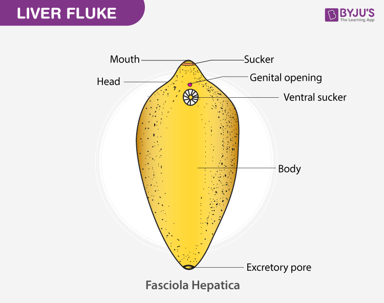
Study Of Various Virtual Specimens Identification With Reasons from cdn1.byjus.com If you live in an area where fluke prevalence is high, speak to your farm vet about forecasting and prevention of transmission. !community rules for liver fluke prevention and control, monitoring by public health officer, village health volunteer, 1 time/week. Radiolucent shadows of flukes may be seen by cholangiography. Deeply culturally rooted habits of raw and undercooked fish dishes consumption are the origin of the infection. In this article we will discuss about the external morphology of liver flukes. The most common types of liver flukes are clonorchis sinensis, opisthorchis viverrini and opisthorchis felineus. Learn vocabulary, terms and more with flashcards, games and other study tools. Liver fluke control involves treatment of infected animals, reduction of the.
Diagnosis of liver fluke is not simple.
Recommendations for the control of liver flukes (fasciola hepatica) in cattle are based on strategically timed treatments with flukicidal. They are principally parasites of the liver of various mammals, including humans. Diagnosis of liver fluke is not simple. Ingestion of fresh water plants with metacercaria or by drinking water with floating metacercariae. Other known risk factors for cholangiocarcinoma include hepatitis b, hepatitis c, alcoholic liver disease and other causes of bile duct inflammation. The signs of having a liver. Unlabeled digestive system diagram diagram human digestive system diagram unlabeled. They occur worldwide and range in size from about 5 millimetres (0.2 inch). There are more than 10,000 species of flukes. Ultrasonography and computed tomography are uselirl in the demonstration of lesions in the liver and biliary tracts. Mode of transmission of liver fluke. There there are proper labelling of this diagram. Liver fluke in sheep also known as:
Asian pacific journal of cancer prevention, vol 17, 2016. A liver fluke is a parasitic worm. The diagram illustrates the four year treatment strategy demonstrated by parr and gray (2000) in which. Fluke infection is estimated to cost the uk agriculture industry about £300 million a year. Liver fluke may also be found in irrigation areas.
Dicrocoelium Dendriticum Wasmuth Lab from wasmuthlab.files.wordpress.com A liver fluke (bovine faciolosis) is a parasitic nematode worm that can cause substantial liver damage within its host. Infections in humans usually occur after eating contaminated raw or undercooked freshwater fish or watercress. !community rules for liver fluke prevention and control, monitoring by public health officer, village health volunteer, 1 time/week. A liver fluke is a parasitic worm. Veinous system, arterial system, circulatory system, schema: Felineus and clonorchis sinensis, are highly endemic in many areas in asia and eastern europe. Liver fluke disease is a chronic parasitic disease of the bile ducts. Ultrasonography and computed tomography are uselirl in the demonstration of lesions in the liver and biliary tracts.
Liver fluke disease is a chronic parasitic disease of the bile ducts.
Internal structure of liver fluke with corresponding designations. Liver flukes are an important cause of acute and chronic disease in grazing sheep and cattle. There there are proper labelling of this diagram. Liver fluke may also be found in irrigation areas. Caused by a flat worm called fasciola hepatica. The most common types of liver flukes are clonorchis sinensis, opisthorchis viverrini and opisthorchis felineus. Veinous system, arterial system, circulatory system, schema: Radiolucent shadows of flukes may be seen by cholangiography. Deeply culturally rooted habits of raw and undercooked fish dishes consumption are the origin of the infection. Fasciola hepatica fasciolosis is an economically important and potentially fatal liver fluke in sheep. These risk factors are thought to be more common causes of. Ancient origins articles related to liver fluke in the sections of history, archaeology, human origins, unexplained, artifacts, ancient places and myths and legends. For example, grazing by the most.
A liver fluke (bovine faciolosis) is a parasitic nematode worm that can cause substantial liver damage within its host. Asian pacific journal of cancer prevention, vol 17, 2016. There there are proper labelling of this diagram. Browse and download thousands of. Radiolucent shadows of flukes may be seen by cholangiography.
Clonorchis The Liver Fluke Of Humans Diagram Quizlet from o.quizlet.com The guide includes detailed diagrams of: !community rules for liver fluke prevention and control, monitoring by public health officer, village health volunteer, 1 time/week. Vector illustration in flat style isolated over white background. For example, grazing by the most. Liver flukes are an important cause of acute and chronic disease in grazing sheep and cattle. Caused by a flat worm called fasciola hepatica. Liver fluke control involves treatment of infected animals, reduction of the. How is the disease transmitted and spread?
Radiolucent shadows of flukes may be seen by cholangiography.
The most common types of liver flukes are clonorchis sinensis, opisthorchis viverrini and opisthorchis felineus. Unlabeled digestive system diagram diagram human digestive system diagram unlabeled. Fasciola hepatica (the common liver fluke or sheep liver fluke), which causes fascioliasis and typically infects sheep and cattle. A liver fluke (bovine faciolosis) is a parasitic nematode worm that can cause substantial liver damage within its host. They are principally parasites of the liver of various mammals, including humans. Liver flukes are one of many factors that have been associated with cholangiocarcinoma. Recommendations for the control of liver flukes (fasciola hepatica) in cattle are based on strategically timed treatments with flukicidal. Browse and download thousands of. Other known risk factors for cholangiocarcinoma include hepatitis b, hepatitis c, alcoholic liver disease and other causes of bile duct inflammation. There are more than 10,000 species of flukes. Asian liver fluke infections, namely infection with the trematode helminths opisthorchis viverrini, o. Internal structure of liver fluke with corresponding designations. In the continental u.s., fasciola hepatica blood chemistries suggestive of liver disease and eosinophilia support the diagnosis.
Felineus and clonorchis sinensis, are highly endemic in many areas in asia and eastern europe diagram of liver. Asian pacific journal of cancer prevention, vol 17, 2016.
Share :
Post a Comment
for "Diagram Of Liver Fluke - Liver Fluke Transverse Section Stock Image Z160 0130 Science Photo Library"


Post a Comment for "Diagram Of Liver Fluke - Liver Fluke Transverse Section Stock Image Z160 0130 Science Photo Library"